Paediatric Spirometry
Paediatric Spirometry
Dec 02, 2025
Paediatric Spirometry
Paediatric spirometry is a key lung function test that measures airflow and volume to assess obstruction, restriction, disease progression, and treatment response, using GLI reference standards and strict quality criteria.
 cAPFP
cAPFP
1. What is Spirometry?
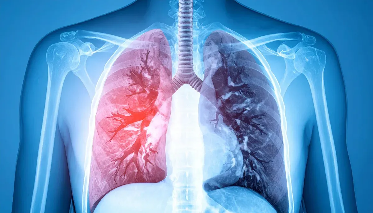
Spirometry is a non-invasive, effort-dependent test that measures airflow and exhaled volume during a maximal forced expiration. It is the most widely used lung function test in children and provides objective measurements such as Forced Vital Capacity (FVC) and Forced Expiratory Volume in 1 second (FEV₁). These are used for early diagnosis, assessing disease severity, monitoring progression and evaluating treatment response.
2. What Does Spirometry Measure?
Spirometry describes how much air a child can move and how quickly, using standardised forced breathing manoeuvres.
2.1 Core Spirometric Indices
- FVC (Forced Vital Capacity) – total volume forcibly exhaled from full inspiration to full expiration. Reduced in restrictive disease and in severe obstruction with air trapping.
- FEV₁ (Forced Expiratory Volume in 1 second) – volume exhaled in the first second of forced expiration. Low values indicate difficulty expelling air quickly due to airway obstruction.
- FEV₁/FVC ratio – proportion of FVC exhaled in the first second. A reduced ratio indicates airflow limitation. In healthy children under 10 years it is usually >0.85; interpretation should use the lower limit of normal (LLN) rather than a fixed cut-off.
- PEF (Peak Expiratory Flow) – maximum flow during forced expiration. Reflects large airway function and effort.
- FEF₂₅–₇₅% (Forced Expiratory Flow 25–75%) – average flow during the middle half of forced expiration. It is sensitive to small airway changes but is effort- and FVC-dependent, so it should not be interpreted in isolation.
2.2 Lung Volumes and Capacities Relevant to Spirometry
- Tidal Volume (VT) – air moved in a normal quiet breath.
- Inspiratory Reserve Volume (IRV) – extra air inhaled after a normal inspiration.
- Expiratory Reserve Volume (ERV) – extra air exhaled after a normal expiration.
- Residual Volume (RV) – air remaining after maximal expiration (not measured by spirometry).
Key capacities:
- Vital Capacity (VC) = IRV + VT + ERV (slowly exhaled = SVC).
- Forced Vital Capacity (FVC) – same volume as VC but exhaled forcefully and rapidly.
- Functional Residual Capacity (FRC) = ERV + RV (not measured by spirometry).
- Total Lung Capacity (TLC) = RV + VC (not measured by spirometry alone).
Spirometry directly measures FVC, FEV₁ and flow indices; TLC, RV and FRC require additional techniques.
3. Indications for Paediatric Spirometry
Spirometry should be requested with a clear clinical question.
| Category | Purpose |
|---|---|
| Diagnosis | Evaluate persistent cough, recurrent wheeze, dyspnoea, exercise limitation; screen high-risk groups (e.g. preterm birth, previous pulmonary TB); pre-operative assessment. |
| Monitoring | Track progression/stability of asthma, cystic fibrosis, post-prematurity or post-TB lung disease; assess treatment or environmental effects. |
| Disability / Impairment | Contribute to rehabilitation, education planning and medico-legal assessments. |
| Other | Epidemiological studies, school or workplace screening, sports/fitness clearance (where appropriate). |
Interpret results using paediatric GLI race-neutral reference equations where available.
4. Contraindications to Paediatric Spirometry
Spirometry is generally safe, but forced expiratory manoeuvres transiently increase intrathoracic, intra-abdominal, intracranial and intra-ocular pressures. There are no absolute contraindications, but the following relative contraindications (adapted from ATS/ERS 2019) should be considered.
| Group | Examples |
|---|---|
| Serious cardiac disease | Recent myocardial infarction (within 1 week); unstable arrhythmia; decompensated heart failure; unstable pulmonary embolism; severe pulmonary hypertension. |
| Recent surgery / neurological risk | Thoracic or abdominal surgery within 4 weeks; brain or eye surgery or known cerebral aneurysm; recent concussion with ongoing symptoms. |
| ENT / middle-ear problems | Sinus or middle-ear infection or surgery within 1 week. |
| Risk from raised intrathoracic/abdominal pressure | Known or suspected pneumothorax; late-term pregnancy in adolescents. |
| Infection control concerns | Suspected or confirmed transmissible respiratory or systemic infection (e.g. TB, influenza, RSV); haemoptysis or copious secretions; oral lesions or active oral bleeding. |
| Syncope / seizures | History of syncope or seizures triggered by cough, straining or forced expiration. |
Stop testing immediately if chest pain, severe breathlessness, dizziness, syncope or marked distress occurs. Do not proceed if the child is crying, in pain, severely anxious or unwilling.
5. Spirometry Pre-test Preparation
5.1 General Preparation
Older children should:
- Avoid smoking, vaping or water-pipe use for at least 1 hour.
- Avoid vigorous exercise for at least 1 hour.
- Avoid intoxicating substances (e.g. alcohol, recreational drugs) for at least 8 hours.
- Wear clothing that does not restrict chest or abdominal movement.
All children:
- Should be calm, seated safely and understand the task.
- May need loose dentures or braces adjusted if they interfere with the mouthpiece seal.
5.2 Bronchodilator Withholding
Where baseline values are required, withhold bronchodilators according to local policy (typical guidance):
- Short-acting β₂-agonists: withhold for 4–6 hours.
- Long-acting β₂-agonists: withhold for 12–24 hours.
- Short-acting anticholinergics: withhold for 6 hours.
Always record:
- Which bronchodilator was used.
- Dose and route.
- Time of last dose.
5.3 Young or Developmentally Delayed Children
In very young children (<5–6 years) or those with developmental delay:
- Attempt spirometry only when clearly indicated.
- Use trained staff, child-friendly equipment and visual incentives.
- Stop if the child is distressed or unable to understand the task.
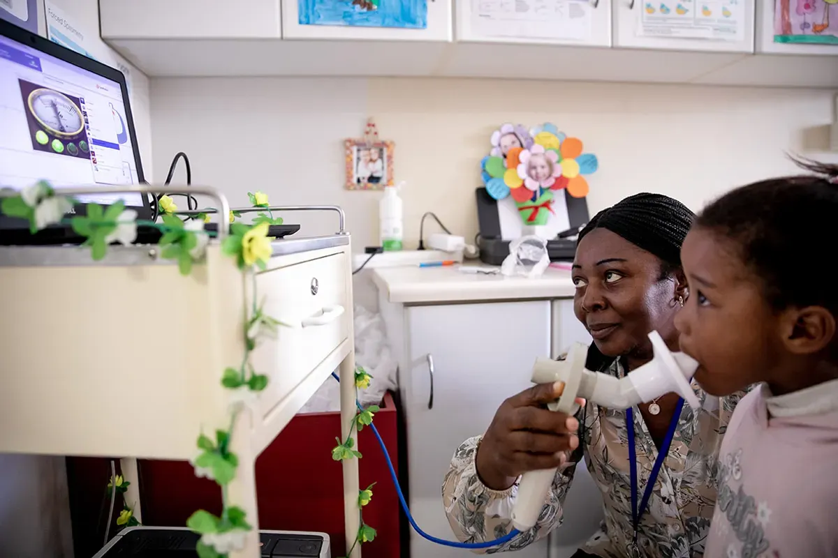
6. Performing Spirometry in Children
The standard FVC manoeuvre has four phases:
- Maximal inspiration to total lung capacity (TLC).
- Sharp, explosive expiration (“blast”) to start the blow.
- Continued forced expiration until no more air can be expelled.
- Rapid full inspiration back to TLC (for flow-volume loops).
6.1 Key Steps
- Explain and demonstrate the manoeuvre in child-friendly language.
- Seat the child upright with feet supported; apply a nose clip.
- Ensure a tight seal around the mouthpiece with tongue flat and inside the teeth.
- Coach a quick, deep breath in to full inspiration.
- Immediately encourage a hard, fast “blast” out and keep blowing to the very end.
- Encourage a rapid full breath back in to complete a flow-volume loop if required.
6.2 Supporting Successful Testing
- Use enthusiastic, consistent coaching and visual incentives.
- Provide rest between blows, especially in younger or sick children.
- Limit attempts according to age, clinical status and local guidance.
7. Evaluating Spirometry Data Quality
Quality assessment includes:
- Acceptability – is each blow technically sound?
- Usability – if not fully acceptable, are FEV₁ / FEV₀.₇₅ / FVC still valid?
- Repeatability – are the best values close together?
- Session quality grading – overall confidence that results reflect the child’s best effort.
7.1 Acceptability Criteria (Single Manoeuvre)
| Component | Children > 6 years | Children ≤ 6 years |
|---|---|---|
| Start of test | - BEV <5% of FVC or <100 mL; rapid rise to peak flow (10–90% rise time ≤150 ms) - hesitation time <2 s. |
BEV <5% of FVC or <80 mL; prompt start with minimal hesitation. |
| During test |
No cough or glottic closure within: - the first 1.0 s for FEV₁ - First (0.75 s for FEV₀.₇₅ in younger children) no leaks, extra breaths, mouthpiece obstruction or tongue occlusion exhalation smooth and continuous without abrupt dips or pauses. |
|
| End of forced expiration | > 6yrs - any of: 1. Volume plateau <25 mL over ≥1 s; 2. FET ≥15 s; or 3. FVC within repeatability tolerance of largest FVC. |
< 6yrs - any of 1. Clear attempt to fully empty lungs; 2. FET ≥1 s; or 3. FVC within repeatability tolerance on ≥2 consistent attempts. A volume plateau is not required. |
| Usability notes | - FEV₁ (or FEV₀.₇₅) may be usable even if EOFE criteria are not met, provided the start and mid-test are acceptable. - If FIVC) – FVC >100 mL or >5%, the manoeuvre is not acceptable. |
|
7.2 Usability Criteria
FEV₁ (or FEV₀.₇₅) and/or FVC are not usable if there is early termination; cough within the first second; major leak or obstruction; or failure to achieve a rapid start.
7.3 Repeatability Criteria (Within Session)
Use only acceptable or usable manoeuvres.
| Age group | Requirements |
|---|---|
| >6 years | Top two FEV₁ values within 150 mL; top two FVC values within 150 mL. |
| ≤6 years | Top two FEV₁ values within 100 mL or 10% of highest value (whichever is greater); same for FVC. |
7.4 Session Quality Grading
Grade (for FEV₁ and FVC separately):
| Grade | Acceptable manoeuvres | Repeatability >6 yrs | Repeatability ≤6 yrs |
|---|---|---|---|
| A | ≥3 | ≤150 mL | ≤100 mL |
| B | 2 | ≤150 mL | ≤100 mL |
| C | ≥2 | ≤200 mL | ≤150 mL |
| D | ≥2 | ≤250 mL | ≤200 mL |
| E | ≥2 or 1 acceptable | >250 mL | >200 mL |
| U | 0 acceptable, >1 usable | — | — |
| F | 0 acceptable, 0 usable | — | — |
8. Interpreting the Spirometry Test Result
8.1 Normal Thresholds
| Parameter | Normal threshold | Notes |
|---|---|---|
| FEV₁, FVC | Z ≥ −1.64 | Within expected range. |
| FEV₁/FVC | Z ≥ −1.64 | LLN, not fixed cut-off; typically >0.85 in children <10 years. |
| FEF₂₅–₇₅% | No fixed LLN | Helpful only when aligned with other indices and curves. |
8.2 Common Spirometric Patterns
| Pattern | FEV₁ | FVC | FEV₁/FVC | Key points |
|---|---|---|---|---|
| Normal | ≥ LLN | ≥ LLN | ≥ LLN | No ventilatory impairment; curves and quality acceptable. |
| Obstructive impairment | ↓ (often more than FVC) | Normal or ↓ | < LLN | Consistent with asthma/airway obstruction; review BDR, curves and exposures. |
| Restrictive (suspected) | Normal or mildly ↓ | ↓ (< LLN) | Normal or ↑ | Suggests reduced lung volume; confirm with TLC where possible. |
| Non-specific/PRISm | ↓ | ↓ | Normal | Could reflect early restriction, early obstruction with air trapping, or poor effort; check quality, consider lung volumes and repeat testing. |
| Physiological dysanapsis | ≥ LLN or mildly ↓ | ≥ LLN | At or just below LLN | Mismatch between airway calibre and lung size; child often asymptomatic; avoid over-diagnosis. |
| Low FEV₁ & FVC from poor effort/muscle weakness | ↓ | ↓ | Normal | Lack of sharp PEF, early termination, inconsistent curves; re-coach; consider neuromuscular weakness if reproducible. |
| Mixed disorder | ↓ | ↓ | < LLN | Features of obstruction and restriction; confirm with full lung function and imaging. |
8.3 Bronchodilator Responsiveness (BDR)
- Repeat spirometry 10–15 minutes after a standard dose of short-acting β₂-agonist.
- Consider a significant BDR when:
- FEV₁ increases by ≥12% and ≥200 mL from baseline, or
- In smaller children, FEV₀.₇₅ or FVC shows similar proportional improvement.
- Interpret BDR in the context of symptoms, baseline lung function and age. A negative BDR does not exclude asthma.
8.4 Grading the Severity of Lung Function Impairment
Use Z-scores:
| Severity | Z-score range | Interpretation |
|---|---|---|
| Normal | ≥ −1.64 | Within expected range. |
| Mild | −1.65 to −2.50 | Mild reduction; may be relevant in context. |
| Moderate | −2.51 to −4.00 | Clear abnormality; likely to impact function. |
| Severe | < −4.00 | Markedly abnormal. |
9. Post-test Procedures
9.1 Recording and Reporting Paediatric Spirometry
- Record demographics (age, sex, height, weight) and reference set used.
- Document pre- and post-bronchodilator status and medications.
- Report best FEV₁, FVC, FEV₁/FVC, PEF and other relevant flows with Z-scores and % predicted.
- Comment on test quality (acceptability, repeatability, overall grade).
9.2 Clinical Management Based on Spirometry Results
- Integrate spirometry with history, examination and other investigations.
- Adjust therapy for asthma or other chronic lung disease according to pattern and severity.
- Use serial measurements to monitor disease control, progression or treatment response.
10. Conclusion
- Spirometry is a cornerstone test for assessing lung function in children.
- Meaningful interpretation requires:
- High-quality, age-appropriate testing technique,
- Use of GLI reference equations and Z-scores,
- Structured quality assessment, and
- Interpretation anchored in clinical context.
Alongside tests such as oscillometry, FeNO and imaging, paediatric spirometry supports early diagnosis, optimised treatment and protection of lung health across childhood.
11. References
- Graham BL, Steenbruggen I, Miller MR, et al. Standardization of Spirometry – 2019 Update. Am J Respir Crit Care Med. 2019;200(8):e70–e88.
- Stanojevic S, Kaminsky DA, Miller MR, et al. ERS/ATS Technical Standard on Interpretive Strategies for Routine Lung Function Tests. Eur Respir J. 2022;60(1):2101499.
- Graham BL, et al. Supplemental Material for: Standardization of Spirometry – 2019 Update. Am J Respir Crit Care Med Online Supplement.